Catalog
Search
43 products
View:
- Selected: 0Areas of use
- Selected: 0Item names
- Selected: 0Manufacturer
- Selected: 0Made in
- Selected: 1Additional
View:
43 products
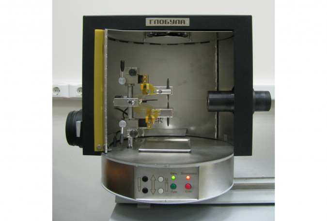
Spectroanalytical tripod "Globula"
The video camera and servos of the electrode holders built into the tripod are designed to automatically maintain the interelectrode gap during the evaporation of the sample by computer analysis in real time of the image of the arc discharge, as well as the initial setting of this gap relative to the optical axis. The electrode holders are cooled with water using a closed-cycle cooling unit.
VMK OPTOELEKTRONIKA
Novosibirsk
Produced in: Novosibirsk
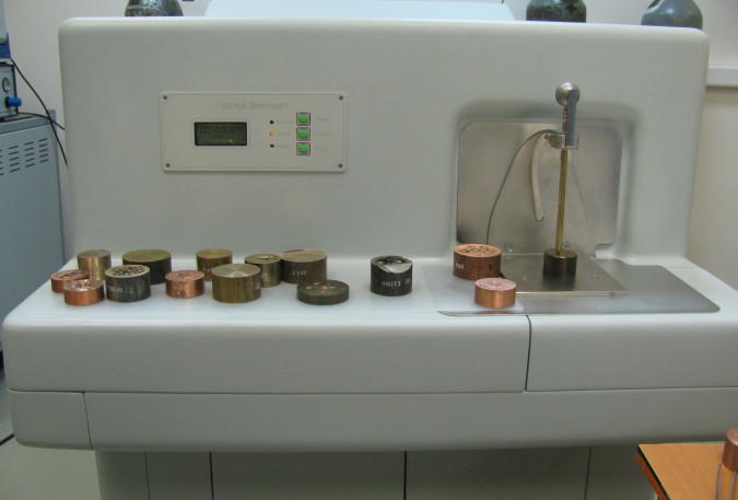
Complex for atomic emission analysis of metals and alloys "Grand Expert"
VMK OPTOELEKTRONIKA
Novosibirsk
Produced in: Novosibirsk
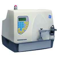
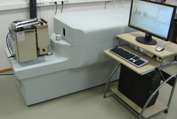
MAX-GF1(2)E-C Spectrometer
The X-ray fluorescence spectrometer SPECTROSCAN MAX-GF1(2)E-C combines two methods of detecting an analytical signal: diffraction on a crystal (wave dispersion - WDX) and energy dispersion (EDX) method, as well as sample delivery adapted for the analysis of large-size images. The collimation of the primary radiation of the X-ray tube and the special design of the sample presentation make it possible to analyze the sample over an area of 1 cm2 in 1 mm increments, and thus investigate the distribution of elements over an area.
The spectrometer is designed to determine the contents of elements in the range from Ca to U in substances in solid, powdery, dissolved states, as well as deposited on the surface or deposited on filters. With the help of fixed energy dispersion channels, any one or two additional elements in the range from magnesium (Mg) to calcium (Ca) can be determined.
With the help of this modification of the spectrometer, forensic and customs examinations, judicial and forensic medical examinations, as well as art-historical examinations are carried out.
NPO Spektron
Saint Petersburg
Produced in: Saint Petersburg
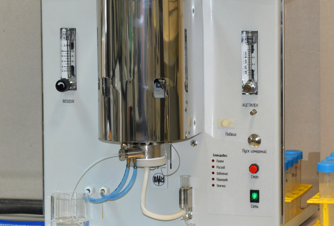
Flame spectrometer "Peacock"
The flame spectrometer is designed for rapid determination of a wide range of concentrations (up to 8 orders of magnitude) of sodium, lithium, potassium, calcium, barium, caesium, rubidium in technological solutions. The excitation of atoms occurs in an air-acetylene flame.
The device consists of a three-slit burner with flame control, a pneumatic sprayer, a spray chamber, an optical radiation input system into the "Hummingbird-2" spectrometer and an automatic air and acetylene supply system, with the possibility to control and adjust gas flow.
The use of a three-slit burner provides increased flame temperature over the central slit of the burner due to the external flame layers. This makes it possible to determine low concentrations of calcium and barium. At the same time, it remains the possibility to determine impurities in highly concentrated solutions without clogging the burner slots.
The lighting system of the spectrometer is mirror-lens, thanks to this, radiation collected from both sides of the burner is introduced into the polychromator.
VMK OPTOELEKTRONIKA
Novosibirsk
Produced in: Novosibirsk
Atomic Emission Complex "Grand Stream"
Atomic Emission Complex "Grand Stream" is designed for rapid determination of the composition of powder samples of natural and industrial origin, includes a "Grand" spectrometer, a "Stream" installation and auxiliary equipment for sample preparation.
VMK OPTOELEKTRONIKA
Novosibirsk
Produced in: Novosibirsk
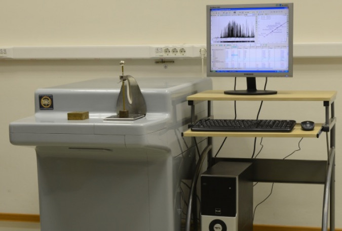
Vacuum Spectrometer Favorite
Vacuum spectrometers are designed for rapid analysis of alloys based on iron, copper, aluminum and other metals in factory and research laboratories, including the determination of elements having analytical lines in the field of vacuum ultraviolet (VUV) (for example, S, P and C in steels).
VMK OPTOELEKTRONIKA
Novosibirsk
Produced in: Novosibirsk
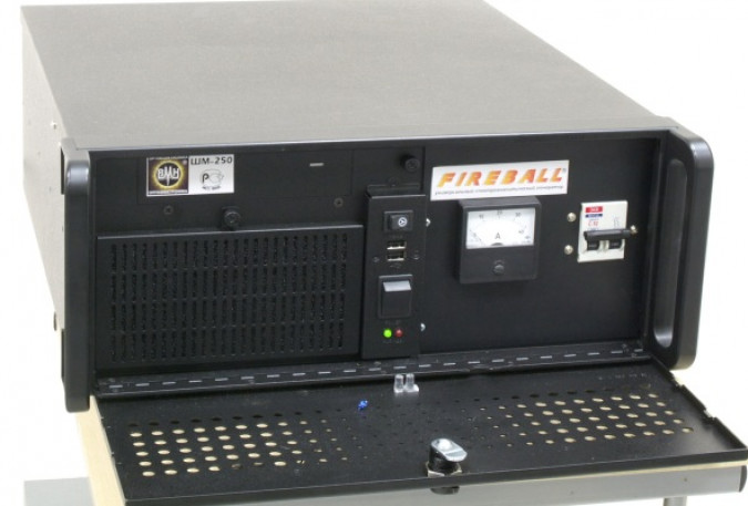
Spectroanalytical generator "Ball lightning"
The generator is designed to produce an electric arc and spark discharge in atomic emission spectral analysis installations. The generator is designed specifically for use in spectral analysis of samples of complex composition for simultaneous determination of impurity and alloying components in a wide range of concentrations.
VMK OPTOELEKTRONIKA
Novosibirsk
Produced in: Novosibirsk

Multichannel analyzer of atomic emission spectra of MAES
The MAES analyzer is a modern means of measuring the intensities of spectral lines and then calculating the concentrations of the elements to be determined.
VMK OPTOELEKTRONIKA
Novosibirsk
Produced in: Novosibirsk
Spectrometer SPECTROSCAN MAX-GVM
The spectrometer is designed to determine the contents of elements in the range from Na to U in substances in solid, powdery, dissolved states, as well as deposited on the surface or deposited on filters.
The principle of operation of the X-ray spectrometer is based on irradiation of the sample with primary radiation from an X-ray tube, measurement of the intensity of secondary fluorescent radiation from the sample at wavelengths corresponding to the elements to be determined, and subsequent calculation of the mass fraction of these elements according to a pre-constructed calibration characteristic, which is the dependence of the content of the element to be determined on the measured intensity.
Secondary fluorescent radiation is decomposed into a spectrum using a crystal analyzer. Due to this, the X-ray fluorescence spectrometer has a high resolution, and therefore the ability to accurately analyze complex multicomponent substances.
NPO Spektron
Saint Petersburg
Produced in: Saint Petersburg
MAES Analyzer for Scintillation
MAES analyzers for recording atomic emission spectra with a time resolution of 1 ms are based on highly sensitive photodiodes and a special switching board for simultaneous reading of several sections of the spectra. The assembly of the rulers is installed on the "Grand" spectrometer.
A " Flow" or twin-jet arc plasmatron is used to excite the atomic emission spectra of powder samples.
The ATOM software registers the selected spectral lines and provides their processing according to the specified algorithms, taking into account the number and brightness of flashes (i.e. particle size).
Recording the spectra of powder samples with a large time resolution makes it possible to estimate the number and size of individual particles, identify their composition and determine the concentrations of elements in each particle, promptly assess the uniformity of the distribution of certain elements to be determined and expand the possibilities of spectral analysis without significant changes in sample preparation.
Main application areas:
mass analysis of powder geological samples by scintillation to determine gold, silver, platinum group metals, iridium, rhodium, etc. with detection limits up to 0.01 g/t;
qualitative determination of the composition of inclusions and mineral particles.
VMK OPTOELEKTRONIKA
Novosibirsk
Produced in: Novosibirsk

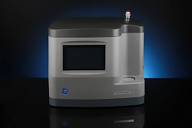
Sulfur Analyzer SPECTROSCAN SW-D3
The analyzer is designed to determine the mass fraction of sulfur in automotive fuel in accordance with GOST R 52660-2006 / GOST ISO 20884-2016, ASTM D6334, as well as in oil and petroleum products according to GOST R 53203-2008 / GOST 33194-2014, ASTM D2622, GB/T 11140-2008. Depending on the customer's needs, the analyzer is configured to work in accordance with a particular standard.
Implements an arbitration method for measuring the mass fraction of sulfur in automobile fuel of the third, fourth and fifth classes.
The device is designed specifically for high-quality and fast X-ray fluorescence analysis of fuel in accordance with the technical regulations "On requirements for automotive and aviation gasoline, diesel and marine fuel, jet fuel and heating oil", which establishes an arbitration X-ray fluorescence wave dispersion method for determining the mass fraction of sulfur in automotive fuels of the third, fourth and fifth classes.
The wave dispersion sulfur analyzer SPECTROSCAN SW-D3 is designed taking into account all the requirements for the process of X-ray fluorescence elemental analysis of low sulfur contents in petroleum products. The operator's actions when performing measurements are minimized:
- the number/name of the sample is entered from the built-in keyboard;
- the sample is poured into two cuvettes;
- the obtained samples are placed in the analyzer and measurements are started.
The analyzer performs all subsequent actions automatically without operator involvement:
- calculates and displays the sulfur content in the sample;
- calculates convergence - the difference in determining the mass fraction of sulfur in the first and second samples;
- prints measurement results on the built-in printer.
NPO Spektron
Saint Petersburg
Produced in: Saint Petersburg
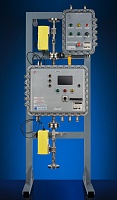
Sulfur Analyzer SPECTROSCAN IS, IS-T
The IS-T and IS X-ray absorption flow SPECTROSCAN analyzers for sulfur in oil are designed to determine the mass fraction of sulfur in the flow of oil/petroleum products in the range from 0.02% to 6.00% (IS-T) or 0.04% to 6.0% (IS).
Analyzers can be used for technological control during transportation and storage of oil, as well as during mixing (compounding) operations. The design of the analyzers allows them to be placed both independently and as part of a unit for measuring oil quality indicators (BIC) or a system for measuring the quantity and quality of oil (CIC). They are available in explosion-proof version.
The analyzers have the function of automatically accounting for the influence of the density of the analyzed medium, as well as the content of water and chloride salts in it on the result of determining the mass fraction of sulfur.
Structurally, the analyzers are a complete product of high factory readiness, having a block-modular design and including all the necessary functional devices to ensure efficient and safe operation.
NPO Spektron
Saint Petersburg
Produced in: Saint Petersburg